Patellar Luxation (Dislocation)
Background
Risk Factors
Patellar luxation is more common in small-breed dogs, with the following more prone to developing the condition:
• Boston terriers
• Chihuahuas
• Miniature poodles
• Pomeranians
• Yorkshire terriers
Certain large-breed dogs may also be affected by a luxating patella, with the following breeds more prone to developing the condition:
• Akitas
• Chinese Shar-Pei
• Flat-Coated retrievers
• Great Pyrenees
Certain cat breeds are also prone to developing the condition:
• Devon Rex
• Abyssinian
Signs
Signs of luxating patella include:
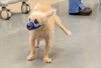
• Lameness in a hind limb
• Skipping gait (holding up hind limb while walking)
Diagnosis
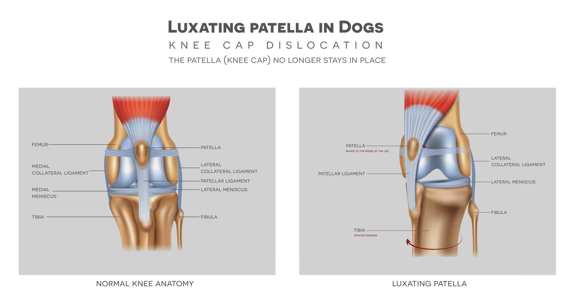
A veterinarian will perform a physical exam of the knee joint to determine if the kneecap is displaced. An x-ray of the hind limb allows the veterinarian to assess the severity of the displacement of the kneecap and any underlying damage to the joint.
Veterinarians grade the luxating patella based on level of severity:
• Grade I – The kneecap can be pushed out of place with the fingers but returns to its normal position when released.
• Grade II – The kneecap occasionally moves out of place spontaneously and can be returned by pushing it back into place with the fingers. Lameness is present.
• Grade III – The kneecap often moves out of place spontaneously and can be pushed back into place with the fingers. Lameness and abnormalities of the hind limb bones are present.
• Grade IV – The kneecap consistently moves out of place and cannot be pushed back into place with the fingers. Lameness and abnormalities of the hind limb bones are severe.
Treatment
Treatments for a luxating patella range from conservative medical management to surgery, depending on the severity of the condition. If surgery is recommended, the specific procedure will depend on the abnormalities causing the kneecap to become displaced. The surgeon may tighten or loosen the soft tissues on either side of the kneecap, deepen the groove in the femur where the kneecap rests, move the tibial crest (where the tendon of the kneecap is attached) to create a better alignment in the leg, or correct any other bone abnormalities present. Surgery may be followed by physical rehabilitation as part of the postoperative treatment plan.
The prognosis for postoperative cases is generally very good. However, pet owners should be on the lookout for concurrent conditions such as a cranial cruciate ligament rupture (equivalent to an ACL rupture in people) or hip dysplasia which may require additional treatment.
Prevention
As this condition is largely hereditary, it cannot be prevented. However, keeping your pet at a healthy weight can reduce complications brought on by this condition.
Make an Appointment
